Diagnosing genetic diseases requires a systematic integration of clinical evaluation, molecular testing, and bioinformatic analysis to identify pathogenic variants responsible for inherited conditions. The core workflow begins with patient phenotyping and family history assessment, followed by selection of the appropriate genomic test (single-gene, panel, exome, or genome sequencing), variant interpretation using established clinical guidelines, and collaborative confirmation through multidisciplinary review. While technological advances have shortened turnaround times to weeks rather than months, the process demands rigorous attention to variant classification standards and quality control at every stage.
The landscape of genetic diagnostics has matured considerably, with next-generation sequencing now serving as a first-tier tool for many clinical scenarios. Healthcare professionals implementing these workflows face practical challenges that extend beyond laboratory technique: selecting the right test for each clinical presentation, navigating insurance authorization, interpreting variants of uncertain significance, and communicating complex results to patients and families. Success depends on understanding not only the molecular methods but also the pre-analytical variables that influence diagnostic yield, from DNA quality to phenotype documentation.
This procedural guide addresses the complete diagnostic pathway from sample collection through clinical reporting. We focus on current best practices for variant interpretation aligned with American College of Medical Genetics and Genomics (ACMG) standards, quality assurance measures to prevent misdiagnosis, and the collaborative frameworks that connect laboratory scientists, genetic counselors, and clinicians. The integration of computational tools with human expertise remains central to accurate diagnosis, particularly as the volume of genomic data continues to expand.
For professionals establishing or refining genetic testing programs, this workflow provides actionable steps grounded in real-world clinical laboratory experience. Ethical considerations, troubleshooting common technical challenges, and verification protocols are embedded throughout to ensure comprehensive coverage of implementation requirements.
Understanding the Foundation: Genomic Approaches to Disease Diagnosis
Genomic approaches to disease diagnosis have fundamentally reshaped clinical practice over the past two decades. What began as labor-intensive single-gene Sanger sequencing, requiring weeks to analyze one candidate gene, has evolved into comprehensive genomic analyses that can interrogate millions of variants across the entire genome in days. This transformation enables clinicians and researchers to diagnose rare genetic disorders that previously remained unidentified, often after years of diagnostic odyssey for patients and families.
The shift reflects three distinct diagnostic modalities now integrated into clinical workflows. Targeted gene panels focus on specific sets of genes associated with particular phenotypes, such as cardiomyopathy panels examining 50-100 cardiac genes or cancer predisposition panels analyzing known hereditary cancer genes. These panels offer cost-effective testing when clinical presentation points to a defined disease category. Whole exome sequencing (WES) captures the protein-coding regions representing approximately 1-2% of the genome but containing roughly 85% of known disease-causing variants. WES has become the workhorse for diagnosing Mendelian disorders, particularly in pediatric and rare disease cases. Whole genome sequencing (WGS) provides the most comprehensive view, detecting variants in coding and non-coding regions, structural variations, and repeat expansions that other methods miss.
Clinical implementation requires understanding when each modality provides diagnostic value. Targeted panels work best when phenotype clearly suggests a gene subset, offering faster turnaround and simpler interpretation. WES suits cases with unclear clinical presentation or suspected rare disorders, balancing breadth with manageable data complexity. WGS addresses cases where WES failed to yield diagnosis, particularly for disorders caused by intronic variants, structural rearrangements, or non-coding regulatory mutations.
The integration of these technologies into clinical practice extends beyond sequencing itself. Laboratories now combine genomic data with phenotype ontologies, variant databases like ClinVar and HGMD, and population frequency resources such as gnomAD to systematically evaluate each variant’s potential pathogenicity. This layered analysis, coupling high-throughput sequencing with sophisticated bioinformatics and clinical correlation, defines contemporary genomic diagnosis, moving the field from hypothesis-driven gene-by-gene testing to unbiased genome-wide investigation.

Tools and Materials Required for Genomic Diagnosis
Sequencing Technologies and Platforms
Next-generation sequencing platforms have become the backbone of genetic disease diagnosis, offering unprecedented throughput and accuracy. Three primary approaches dominate clinical practice in 2026, each suited to different diagnostic scenarios.
Whole genome sequencing analyzes the entire 3 billion base pairs of human DNA, providing comprehensive coverage of coding and non-coding regions. Current platforms achieve 30x coverage or higher, enabling detection of single nucleotide variants, insertions, deletions, and structural variations. This approach excels when phenotypes are non-specific or when regulatory region variants are suspected.
Whole exome sequencing focuses on protein-coding regions, roughly 2% of the genome containing 85% of known disease-causing variants. With faster turnaround times and lower costs than WGS, exome sequencing remains the workhorse for Mendelian disorders where pathogenic variants typically disrupt protein function.
Targeted gene panels sequence predetermined sets of genes associated with specific conditions or phenotypes. Panels range from small sets covering single disorders to comprehensive collections exceeding 500 genes. They offer deeper coverage at specific loci, higher sensitivity for low-level mosaicism, and streamlined interpretation, critical advantages when clinical presentation suggests specific diagnostic categories like cardiomyopathy or hereditary cancer syndromes.
Bioinformatics and Analysis Infrastructure
Genomic diagnosis generates massive datasets, typically 100-200 gigabytes per whole genome sequence, requiring robust computational infrastructure to process and interpret. Clinical laboratories need high-performance computing clusters or cloud-based platforms capable of handling parallel processing workflows that align millions of sequence reads to the reference genome in hours rather than days.
Pipeline software like GATK (Genome Analysis Toolkit), FreeBayes, and Sentieon perform the core tasks of sequence alignment, variant calling, and quality filtering. These tools identify genetic differences between patient samples and reference genomes, flagging approximately 3-4 million variants per whole genome that require systematic review.
Variant interpretation relies heavily on curated databases. ClinVar aggregates clinical significance classifications from laboratories worldwide, while gnomAD provides population frequency data essential for distinguishing pathogenic mutations from benign polymorphisms. OMIM catalogs gene-disease relationships, and HGMD documents published disease-causing mutations.
Clinical decision support systems like Fabric Genomics or Genomenon integrate multiple data sources, applying ACMG-AMP guidelines to prioritize variants for manual review. These platforms reduce interpretation time from weeks to hours by automatically classifying variants based on established criteria, though expert oversight remains critical for complex cases.
Quality Control and Validation Resources
“`
Quality control and validation resources form the backbone of reliable genetic disease diagnosis, ensuring that results meet clinical-grade accuracy standards. Laboratories conducting diagnostic genomics require a comprehensive suite of reference materials that serve as benchmarks throughout the testing process.
Certified reference materials from organizations like the Genome in a Bottle Consortium (GIAB) provide validated DNA samples with known variant profiles across the genome. These materials enable laboratories to assess the sensitivity and specificity of their sequencing and bioinformatics pipelines, identifying any systematic errors or detection gaps. Clinical laboratories typically run these controls alongside patient samples in every sequencing batch to verify platform performance.
Proficiency testing programs, such as those offered by the College of American Pathologists (CAP), deliver blind samples with unknown genotypes that laboratories must correctly identify to maintain accreditation. These external quality assessments validate the entire diagnostic workflow from sample processing through variant interpretation.
Validation protocols must be established for each gene or panel before clinical use, documenting the test’s analytical sensitivity, specificity, positive predictive value, and limit of detection. This includes demonstrating consistent performance across variant types, single nucleotide variants, insertions, deletions, and copy number changes, and challenging genomic regions with high GC content or repetitive sequences.

Safety Considerations and Ethical Safeguards
Laboratory Safety and Sample Handling
Genetic disease diagnosis begins with rigorous sample handling protocols that protect both laboratory personnel and specimen integrity. All biological materials require BSL-2 containment practices at minimum, work in certified biosafety cabinets, wear appropriate personal protective equipment including lab coats and gloves, and follow institutional bloodborne pathogen exposure plans.
Contamination represents the greatest threat to diagnostic accuracy. Implement strict DNA-free workflow zones, use barrier-tipped pipettes exclusively, and maintain separate pre- and post-PCR areas with dedicated equipment. Never process high-concentration amplicons near patient samples. Change gloves between specimens, decontaminate work surfaces with 10% bleach solution, and UV-irradiate hoods before each session.
Sample tracking demands meticulous chain-of-custody documentation. Label specimens with at least two unique identifiers, log collection timestamps, and maintain refrigeration at 2-8°C for DNA extraction within 24 hours or freeze at -80°C for long-term storage. Document freeze-thaw cycles, each degrades DNA quality. Verify sample identity through barcode scanning at every transfer point to prevent potentially catastrophic diagnostic misattribution.
Data Security and Patient Privacy
Genomic data requires stringent security measures because it contains immutable personal identifiers that can reveal disease risks, ancestry, and familial relationships. Healthcare organizations must implement encryption both for data in transit and at rest, meeting HIPAA encryption requirements that mandate AES-256 or equivalent standards for stored genomic files. Secure socket layer (SSL) protocols protect data transmission between sequencing platforms, analysis servers, and clinical databases.
Access controls are critical: role-based permissions ensure that only authorized personnel can view genomic data, with audit logs tracking every access event. Two-factor authentication should be mandatory for all systems handling genetic information. Cloud storage solutions must be HIPAA-compliant, with business associate agreements in place and servers located within jurisdictions that respect patient privacy laws.
De-identification presents unique challenges for genomic data, since even without traditional identifiers, genetic sequences can potentially be re-identified. Storage systems should separate genomic data from demographic information, using secure tokens to link them only when clinically necessary. Regular security audits and penetration testing help identify vulnerabilities before breaches occur.
Ethical Considerations in Genetic Testing
Obtaining truly informed consent for genomic testing requires explaining not just what the test will look for, but what it might uncover beyond the initial clinical question. Patients must understand the possibility of incidental findings, pathogenic variants unrelated to their presenting condition, and decide in advance which categories of secondary findings they wish to receive. The American College of Medical Genetics and Genomics recommends offering analysis of actionable secondary findings in at least 81 genes associated with medically important conditions, but patients retain the right to decline this information.
Genetic counseling before and after testing is essential. Counselors help patients comprehend inheritance patterns, recurrence risks, and implications for family members. They navigate the psychological impact of learning carrier status or predisposition to late-onset diseases, particularly when no treatment exists.
Managing variants of uncertain significance presents ethical complexity. These findings cannot confirm or rule out disease, yet withholding them entirely may seem paternalistic. Clear communication about their limitations prevents misinterpretation while preserving diagnostic transparency. Documentation of patient preferences regarding recontact when variant classifications change ensures respect for autonomy throughout the diagnostic journey.
Step-by-Step Diagnostic Process
Step 1: Clinical Assessment and Test Selection
The diagnostic journey begins with a thorough clinical assessment that determines which genomic test will yield the most clinically actionable results. Clinicians collect a detailed patient and family history, documenting symptoms, age of onset, disease progression, and any relevant environmental exposures. This information establishes the phenotypic profile that guides subsequent testing decisions.
Phenotypic analysis involves systematic evaluation of physical findings, laboratory abnormalities, and imaging results that may suggest a genetic etiology. Healthcare professionals look for patterns that match known genetic syndromes, such as developmental delays paired with dysmorphic features, or multisystem involvement that points to metabolic disorders. Detailed photographic documentation and anthropometric measurements provide objective data for correlation with genetic findings.
Constructing a three-generation family pedigree reveals inheritance patterns, affected relatives, and consanguinity that influence test selection. A clear autosomal dominant pattern with complete penetrance may warrant targeted gene panel testing, while complex presentations with unclear inheritance suggest whole exome or genome sequencing.
Test selection balances diagnostic yield against cost and turnaround time. Single-gene testing suits well-characterized conditions with pathognomonic features. Gene panels work for phenotypically heterogeneous disorders like cardiomyopathies or hereditary cancer syndromes. Exome sequencing addresses atypical presentations or diagnostic odysseys where targeted approaches failed. Genome sequencing captures structural variants and noncoding regions when comprehensive analysis is needed.

Step 2: Sample Collection and Preparation
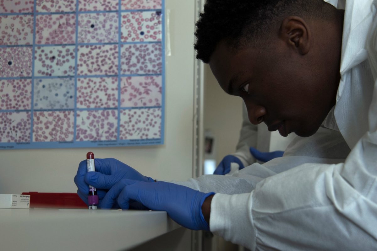
Specimen quality determines downstream diagnostic accuracy, making proper collection technique essential. For constitutional genetic testing, peripheral blood collected in EDTA tubes remains the gold standard, providing high-quality, high-molecular-weight DNA suitable for all sequencing platforms. Saliva samples offer a non-invasive alternative, though DNA yield and quality vary more than blood. Tissue biopsies become necessary for tumor genomic profiling or when mosaicism is suspected, requiring fresh-frozen or formalin-fixed paraffin-embedded preservation depending on analysis needs.
DNA extraction follows standardized protocols using commercial kits that ensure consistent purity and concentration. Quality assessment measures DNA integrity through gel electrophoresis or automated systems, checking for degradation that compromises sequencing. Spectrophotometry confirms concentration and identifies protein or phenol contamination through absorbance ratios. For whole genome sequencing, DNA quantity requirements typically range from 200 nanograms to 1 microgram.
Library preparation involves fragmenting DNA to appropriate sizes, attaching sequencing adapters, and adding sample-specific barcodes for multiplexing. Automated liquid handling systems reduce variability, while quality control metrics verify proper fragment size distribution and adapter ligation efficiency before committing samples to expensive sequencing runs.

Step 3: Sequencing and Data Generation
With sequencing libraries prepared, the next critical phase involves executing the sequencing run and monitoring real-time quality metrics to ensure data integrity. Modern sequencing platforms generate quality scores throughout the process, with platforms like Illumina systems providing cluster density metrics, phasing values, and Q-scores that should be monitored during each cycle.
For whole genome sequencing, target coverage depth typically ranges from 30×-40× for clinical applications, while whole exome sequencing aims for 100×-150× coverage across targeted regions. Targeted panels may require 500× or higher coverage depending on the application. During the run, operators monitor cluster generation efficiency (ideally 80-90%), template generation rates, and error rates per cycle.
Initial data output validation begins immediately after sequencing completion. Assess overall yield, the percentage of bases exceeding Q30 quality scores (target ≥75%), and read length distribution. Generate preliminary FastQC reports to identify adapter contamination, GC bias, or per-base quality degradation. These metrics determine whether the run meets specifications for downstream analysis or requires troubleshooting and potential re-sequencing before proceeding to variant calling.
Step 4: Bioinformatics Analysis and Variant Calling
Sequence alignment serves as the first critical bioinformatics step, mapping raw sequencing reads to the human reference genome (GRCh38/hg38). Alignment algorithms like BWA-MEM or Bowtie2 position each short DNA fragment to its corresponding genomic location, accounting for sequencing errors and legitimate genetic variation. Proper alignment quality is essential, misaligned reads lead directly to false variant calls.
Variant calling algorithms then identify positions where the patient’s sequence differs from the reference genome. Tools such as GATK HaplotypeCaller, DeepVariant, or FreeBayes detect single nucleotide variants, insertions, deletions, and structural variations. Modern callers use sophisticated statistical models to distinguish true variants from sequencing artifacts, considering read depth, base quality scores, and strand bias. For clinical applications, you need minimum coverage thresholds, typically 20x for heterozygous variants and 10x for homozygous calls.
Filtering criteria eliminate low-quality calls and common population variants unlikely to cause disease. Initial filters remove variants with inadequate read depth, poor mapping quality, or excessive strand bias. Population frequency filters flag variants present above specific thresholds in databases like gnomAD, since common variants rarely cause rare diseases.
Annotation enriches remaining variants with biological context using tools like VEP or ANNOVAR. This process adds gene names, predicted protein consequences, conservation scores, existing clinical interpretations from ClinVar, and functional predictions from algorithms like SIFT or PolyPhen-2, preparing variants for clinical interpretation.
Step 5: Clinical Interpretation of Variants
Once variant calling is complete, you must evaluate which variants are clinically relevant to your patient’s presentation. This interpretation step determines whether identified variants explain the observed phenotype and requires systematic application of standardized classification frameworks.
Begin by applying the ACMG-AMP variant guidelines to classify each candidate variant. These criteria organize evidence into pathogenic, likely pathogenic, uncertain significance, likely benign, and benign categories using specific evidence types. Population frequency data serves as your first filter, variants present above 5% in healthy populations rarely cause Mendelian disease. Examine computational predictions from tools like SIFT, PolyPhen-2, and CADD scores, though these provide supporting rather than standalone evidence.
Critically assess variant-phenotype correlation by comparing your patient’s clinical features against published reports in ClinVar, OMIM, and disease-specific databases. Does the variant occur in a gene definitively linked to the patient’s symptoms? Match the inheritance pattern you observe in the family pedigree to the gene’s known mechanism. A heterozygous variant in a recessive disease gene typically won’t explain the phenotype unless you identify a second pathogenic allele.
Functional impact determines pathogenicity for many variants. Nonsense and frameshift variants in critical exons generally disrupt protein function, while missense variants require deeper evaluation. Review whether the variant affects conserved residues, disrupts protein domains, or has demonstrated functional consequences in laboratory studies.
Document your interpretation rationale systematically, noting which ACMG criteria apply and their strength of evidence.
Step 6: Multidisciplinary Review and Validation
Genetic disease diagnosis rarely occurs in isolation. Once bioinformatics analysis identifies potentially causative variants, assembling a multidisciplinary team ensures comprehensive evaluation before finalizing clinical reports. This collaborative review typically includes medical geneticists, the ordering physician, genetic counselors, bioinformaticians, and relevant subspecialists based on the patient’s presentation.
Regular molecular case conferences provide structured forums for discussing complex cases. During these meetings, teams review phenotype-genotype correlations, assess whether identified variants explain the full clinical picture, and determine if additional testing is warranted. Genetic counselors contribute essential context about family history and inheritance patterns that may influence variant interpretation.
For variants with uncertain clinical significance or unexpected findings, confirmatory testing becomes necessary. This might involve Sanger sequencing to validate specific variants, parental testing to establish de novo status, or RNA studies to assess splicing effects. Complex cases may require consultation with external experts or submission to ClinGen expert panels.
Document all review discussions, dissenting opinions, and the rationale behind final classifications. This transparency strengthens diagnostic confidence and provides valuable context for future reanalysis as genomic knowledge evolves.
Step 7: Diagnostic Reporting and Clinical Communication
The final diagnostic report serves as the critical bridge between genomic analysis and patient care. Structure reports following standardized frameworks that include patient demographics, test methodology, variant classification using ACMG-AMP terminology, clinical interpretation, and actionable recommendations. Present findings hierarchically: lead with definitive pathogenic variants, followed by likely pathogenic variants, then variants of uncertain significance if clinically relevant.
Separate communications for ordering clinicians and patients. Clinician reports should contain technical details about variant nomenclature, population frequencies, functional predictions, and evidence supporting pathogenicity classifications. Include differential diagnoses when multiple conditions could explain findings. Patient-directed summaries require plain language explanations, focusing on health implications rather than molecular details.
Coordinate with genetic counselors before patient disclosure, particularly for unexpected findings or uncertain results. Specify recommended clinical actions: surveillance protocols, therapeutic interventions, family cascade testing, or periodic reassessment timelines. When reporting negative results, clarify test limitations and residual diagnostic possibilities. Document variant reclassification pathways since interpretations may change as genomic knowledge advances. Ensure reports meet regulatory requirements for clinical laboratories while remaining comprehensible to multidisciplinary care teams.
Verification and Confirmation Methods
Sanger Sequencing Confirmation
Sanger sequencing remains the gold standard for confirming pathogenic variants identified through next-generation sequencing platforms. Clinical laboratories should perform Sanger confirmation for all reportable pathogenic or likely pathogenic variants before releasing diagnostic reports, as this orthogonal method validates both the variant call and its zygosity status.
The confirmation process involves designing primers flanking the variant of interest, typically 300-500 base pairs upstream and downstream. Amplify the target region using PCR, purify the product, and sequence bidirectionally to eliminate strand-specific artifacts. Compare the resulting chromatogram against the reference sequence to verify the exact nucleotide change and assess whether the variant appears heterozygous or homozygous.
Sanger confirmation proves particularly critical when NGS coverage falls below 30x at the variant position, when the variant occurs in repetitive or GC-rich regions prone to sequencing errors, or when clinical management decisions hinge on the finding. While Sanger sequencing cannot replace comprehensive genomic analysis, it provides essential validation that protects against false positive reports and ensures diagnostic accuracy in clinical decision-making.
Familial Segregation Analysis
Familial segregation analysis examines whether a variant co-segregates with disease across multiple family members, providing crucial evidence for pathogenicity assessment. When you identify a candidate variant in a proband, testing affected and unaffected relatives reveals whether the variant tracks with the phenotype as predicted by Mendelian inheritance patterns.
For dominant conditions, the variant should appear in all affected individuals and absent in unaffected family members (accounting for incomplete penetrance). Recessive disorders require demonstrating that affected individuals carry biallelic variants while unaffected parents are heterozygous carriers. De novo variants gain support when confirmed absent in both unaffected parents, though you must account for the possibility of germline mosaicism.
The strength of segregation evidence depends on family structure and size. A variant segregating with disease across three or more affected relatives in multiple generations provides strong supporting evidence under ACMG-AMP criteria. Conversely, finding the variant in unaffected relatives (particularly older individuals past typical disease onset) argues against pathogenicity.
Document each tested relative’s phenotype carefully, including age and clinical details, as this context determines whether segregation data truly supports or refutes causality.
Functional Validation Approaches
When clinical databases and computational predictions yield inconclusive results for a detected variant, functional studies provide experimental evidence of its biological impact. These laboratory techniques are particularly valuable for novel missense variants in genes with limited phenotypic data or when family segregation analysis is unavailable.
In vitro expression assays measure whether a variant alters protein production levels or stability. Researchers introduce the wild-type and mutant sequences into cell lines, then quantify protein expression through Western blotting or immunofluorescence. A significant reduction in protein levels suggests a loss-of-function mechanism.
Enzyme activity assays directly test whether variants affecting metabolic genes impair catalytic function. Comparing substrate conversion rates between normal and variant proteins reveals functional deficits that correlate with disease severity.
RNA splicing analysis examines whether intronic or exonic variants create cryptic splice sites or disrupt normal splicing patterns. RT-PCR followed by sequencing of cDNA reveals aberrant transcripts that escape in silico prediction tools.
For variants predicted to affect protein-protein interactions, yeast two-hybrid systems or co-immunoprecipitation experiments demonstrate whether binding capacity is disrupted. These assays are especially relevant for variants in multi-subunit complexes or signaling pathways where interaction integrity determines function.
Common Challenges and Troubleshooting
Genomic diagnosis inevitably confronts healthcare professionals with three persistent challenges: variants of uncertain significance (VUS), technical limitations inherent to sequencing platforms, and interpretation complexities arising from incomplete reference data. Understanding how to address these obstacles systematically improves diagnostic yield and prevents misdiagnosis.
VUS represent the most common diagnostic challenge, accounting for approximately 15-30% of clinical sequencing results depending on the gene panel used. When encountering a VUS in a gene strongly associated with the patient’s phenotype, pursue segregation analysis in affected and unaffected family members. If the variant segregates perfectly with disease and is absent in unaffected relatives, this evidence can reclassify the variant toward likely pathogenic status. Conversely, presence in healthy individuals suggests benign classification.
Access to functional studies provides another route when clinical evidence remains insufficient. Submit candidate variants to model organism databases or specialized laboratories offering functional assays, though recognize that turnaround times typically span months rather than weeks.
Technical limitations present differently depending on sequencing modality. Short-read sequencing struggles with repetitive genomic regions, homopolymers, and structural variants larger than 1-2 kilobases. If clinical suspicion remains high despite negative exome results, consider long-read sequencing platforms or targeted PCR-based methods for problematic regions. Incomplete coverage deserves particular attention, always review coverage depth reports to identify genes or exons that failed to meet quality thresholds, as these represent diagnostic blind spots.
Interpretation complexities often stem from phenotypic heterogeneity and incomplete penetrance. A patient presenting with atypical features for a suspected genetic syndrome may carry a causative variant that existing literature hasn’t yet connected to that phenotype. Maintain diagnostic humility in such cases, clearly documenting limitations in the clinical report rather than forcing interpretation. When genes lack robust clinical evidence, particularly for recently discovered disease associations, weight external evidence cautiously and consider deferring definitive interpretation until additional cases emerge.
For copy number variants near the limits of detection, orthogonal confirmation via chromosomal microarray or multiplex ligation-dependent probe amplification resolves ambiguity more reliably than relying on exome read-depth algorithms alone.
Frequently Asked Questions About Genetic Disease Diagnosis
Implementing genomic diagnosis raises practical questions that directly impact clinical workflows and patient care. These frequently asked questions address the operational realities that healthcare teams encounter when integrating genomic technologies into diagnostic practice.
What diagnostic accuracy can we expect from whole exome sequencing?
Diagnostic yields for whole exome sequencing typically range from 25-50% depending on the clinical phenotype, with higher yields in cases with clear Mendelian inheritance patterns and consanguinity. The actual diagnostic rate varies significantly based on patient selection, phenotypic specificity, and the clinical expertise applied during variant interpretation.
How long does the complete diagnostic process take from sample collection to final report?
Standard genomic diagnostic workflows typically require 4-8 weeks for completion, though this timeline varies by laboratory capacity, test complexity, and whether additional validation or familial studies are needed. Rapid genomic sequencing protocols designed for critically ill patients can deliver preliminary results within 24-72 hours when clinically indicated.
What factors influence insurance reimbursement for genomic testing?
Coverage decisions depend on medical necessity documentation, prior authorization requirements, the specific clinical indication, and whether the test meets payer criteria for diagnostic utility. Comprehensive pre-authorization with detailed clinical justification, family history, and prior testing results significantly improves approval rates.
When should we consider retesting patients who received negative results?
Retesting becomes appropriate when new family members develop similar symptoms, the patient’s phenotype evolves or becomes more specific, significant time has passed allowing for database expansion and improved interpretation methods, or when newer genomic technologies with better coverage become available. Many previously undiagnosed cases achieve diagnosis upon reanalysis as variant databases expand and interpretation guidelines improve.
Beyond these operational considerations, healthcare teams should establish clear protocols for result disclosure and genetic counseling integration. The turnaround time for genomic diagnosis makes pre-test counseling particularly important, as families need realistic expectations about both the timeline and the possibility of uncertain or negative results. Building relationships with genetic counselors before ordering tests streamlines the entire workflow and ensures appropriate patient support throughout the diagnostic journey.
Cost considerations extend beyond the test itself to include the infrastructure required for interpretation, the time commitment from multidisciplinary team members, and the potential need for follow-up testing in relatives. Many institutions find that centralizing genomic expertise within molecular tumor boards or genomics consult services improves both efficiency and diagnostic accuracy while reducing redundant testing. This collaborative model distributes the interpretation workload and ensures that complex cases receive adequate attention from clinicians with relevant subspecialty knowledge.
Diagnosing genetic diseases through genomic technologies represents a fundamentally collaborative endeavor that bridges clinical acumen with cutting-edge scientific innovation. The workflow outlined in this guide demonstrates that successful diagnosis depends not on technology alone, but on the thoughtful integration of sequencing platforms, bioinformatics expertise, clinical interpretation, and multidisciplinary review. Each step requires healthcare professionals to balance technical precision with nuanced clinical judgment, ensuring that genomic data translates into actionable diagnostic insights.
The field continues to evolve rapidly. As sequencing costs decline and analytical algorithms improve, genomic diagnosis becomes increasingly accessible across healthcare settings. New variant databases expand our interpretive capacity, while machine learning tools refine pathogenicity prediction. These advances don’t diminish the need for clinical expertise; rather, they amplify its importance. Clinicians who understand both the capabilities and limitations of genomic technologies can better select appropriate tests, interpret complex results, and communicate findings effectively to patients and families.
Looking forward, the integration of genomic research into routine clinical practice will accelerate diagnosis for rare diseases, enable precision treatment strategies, and uncover previously unrecognized genetic conditions. Success in this landscape requires ongoing education, adherence to evolving diagnostic standards, and commitment to ethical frameworks that protect patient privacy while advancing scientific knowledge. For healthcare professionals and researchers, mastering this diagnostic workflow positions them at the forefront of personalized medicine, where genomic insights drive improved outcomes for patients with genetic diseases.